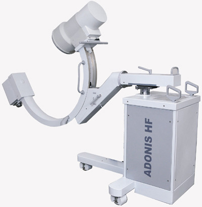
Mobile Fluoroscopic System “Portable C-arm”
C-arm of a mobile X-ray image intensifier
General configuration and range of movements
A mobile image intensifier generally consists of two units, the X-ray generator and image system on a portable imaging system (C-arm) and the workstation unit used to store and manipulate the images. The imaging system unit can perform a variety of movements that allow for use in a variety of surgical procedures such as cardiology, orthopedics and urology. This unit provides the appropriate structure to mount an image intensifier and an X-ray tube with a beam limiting device positioned directly opposite from and aligned centrally to each other.
The C-arm is capable of many movements: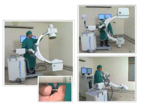
- Horizontal travel: about 200 mm
- Orbital travel: about 115 degrees
- Motorized vertical travel: 460 mm
- Wig-wag about +/-12 cm (entire C-arm and Image Intensifier)
- C-arm rotation about the horizontal axis +/- 210 degrees
The X-ray generator, dose control system and collimator controls are usually housed in the chassis on which the C-arm is mounted. All of the control systems are closed loop systems which are directed by the master controller initial program settings. The master controller generally is found in the work station. User controls on the C-arm allow the operator to modify the operation of the system while in use. I.e. format size, slot collimator position, dose rate etc.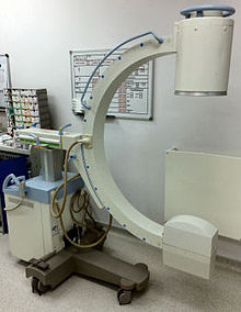
The Imaging system must be compact and lightweight to allow easy positioning with adequate space to work around and a wide range of motion while remaining inflexible enough so as to avoid misalignment due to flexion caused by the mass of the X-ray tube or Image system assemblies