Echocardiogram
 An echocardiogram is an ultrasound examination of the heart. It is a non- invasive test, excellent for assessing the size of heart chambers, valves, “hole-in-the-heart”, presence of clots or tumours, contracting and relaxing of the heart muscle and the efficiency of the heart pump. It cannot, however, visualize blockages in the arteries.
A measurement called the ejection fraction (EF) is measured from the echocardiogram. The prognosis of any heart condition is strongly influenced by this measurement. If the EF is low, the chance of sudden cardiac death is higher. An EF of less than 35% is used as a criteria for implanting an implantable cardioverter defibrillator (ICD).
An echocardiogram is used to investigate heart murmurs. A heart murmur may be “innocent” or due to pathology such as a narrowed or leaky valve, or a hole in the heart. An echocardiogram is able to distinguish between all these causes.
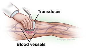 Ultrasound is a study that uses sound waves to “see” inside your body. A carotid duplex ultrasound is performed to evaluate symptoms including dizziness, loss of memory, stroke, loss of musclecontrol and other symptoms that might result from narrowing or blockage of the vessels (carotid arteries) on either side of your neck. At the S. Mark Taper Foundation Imaging Center, we have a specialized team of physicians, nurses, and technologists who are experts in ultrasound radiology.