2D, 3D and 4D Ultrasounds
What are 2D, 3D and 4D ultrasound scans?
3D scans show still pictures of your baby in three dimensions. 4D scans show moving 3D images of your baby, with time being the fourth dimension.
It’s natural to be really excited by the prospect of your first scan. But some mums find the standard 2D scans disappointing when all they see is a grey, blurry outline. This is because the scan sees right through your baby, so the photos show her internal organs.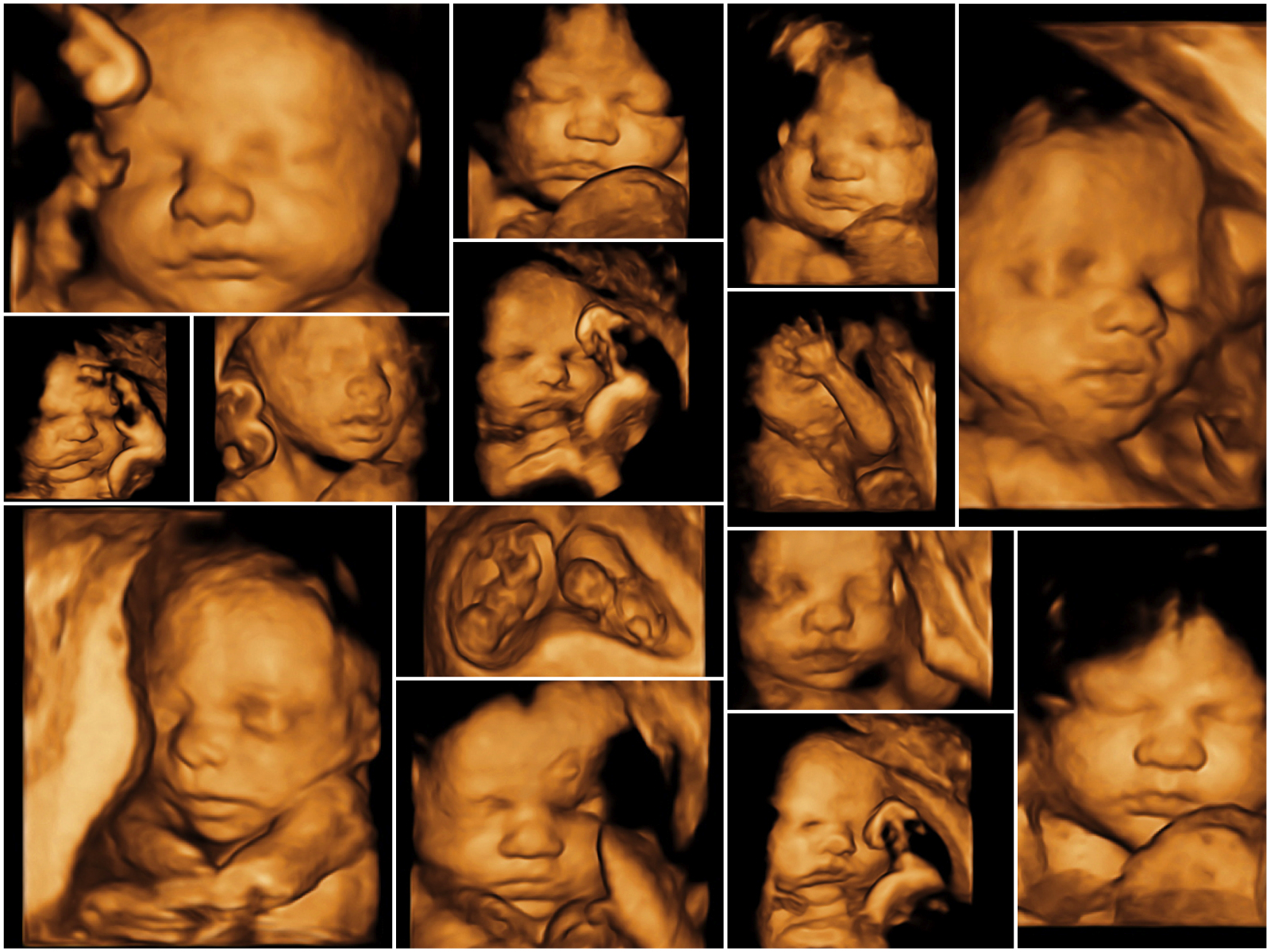
With 3D and 4D scans, you see your baby’s skin rather than her insides. You may see the shape of your baby’s mouth and nose, or be able to spot her yawning or sticking her tongue out.
3D and 4D scans are considered as safe as 2D scans, because the images are made up of sections of two-dimensional images converted into a picture. However, experts do not recommend having 3D or 4D scans purely for a souvenir photo or recording, because it means that you are exposing your baby to more ultrasound than is medically necessary. Some private ultrasounds can be as long as 45 minutes to an hour, which may be longer than recommended safety limits.
3D and 4D scans may nonetheless provide more information about a known abnormality. Because these scans can show more detail from different angles, they can help in the diagnosis of cleft lip. This can help doctors to plan the repair after birth.
3D scanning can also be useful to look at the heart and other internal organs. As a result, some fetal medicine units do use 3D scans, but only when they’re medically necessary.
There’s no evidence to suggest that the scans aren’t safe, and most mums-to-be gain reassurance from them. Nonetheless, any type of ultrasound scan should only be performed by a trained professional, for as short a time and at the lowest intensity, as possible.
If you’d like a 3D or 4D scan you’ll probably need to arrange it privately, and pay a fee. The clinic may also give you a recording of the scan on DVD, though this is likely to cost extra.
The special transducers and software required to do 3D and 4D scans are expensive. There are few clear medical benefits, and experts say they should only be done if there’s a medical need. So it’s unlikely that these scans will replace normal 2D scans used for routine maternity care in the NHS.
If you decide to have one, the best time to have a 3D or 4D scan is when you’re between 26 weeks and 30 weeks pregnant.
Before 26 weeks your baby has very little fat under her skin, so the bones of her face will show through. After 30 weeks, your baby’s head may go deep down in your pelvis, so you may not be able to see her face.
If the placenta is at the front of your womb (uterus), known as anterior placenta, you’ll get the best images of your baby if you wait until 28 weeks.
It’s natural that you’d like to see your baby’s face on the scan. But sometimes it’s not possible, depending on how she’s lying.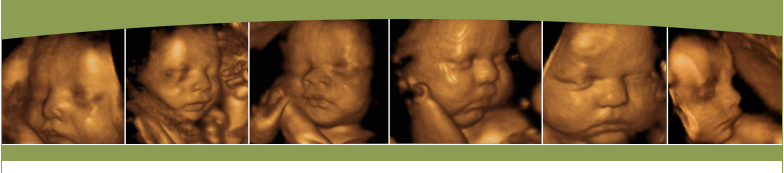
If she’s lying facing outwards, with a good pool of amniotic fluid around her features, you should be able to see her face clearly. But if she’s facing your back, her head’s far down in your pelvis, or there’s not much fluid around her, you won’t see much. The same applies if you have a lot of tummy fat.
The sonographer may ask you to go for a walk, or to come back in a week, when your baby may have moved to a better position. If it’s not possible to get good views of her face, you may be able to see her fingers and toes instead.
Some private providers may offer you a free repeat scan if you can’t see your baby’s face. However, others will advise you of the limits of ultrasound and may not offer to repeat the scan. Check the company’s policy before parting with any money.